Cardiac action potentials[edit]Main articles: Cardiac action potential, Electrical conduction system of the heart, Cardiac pacemaker and Arrhythmia
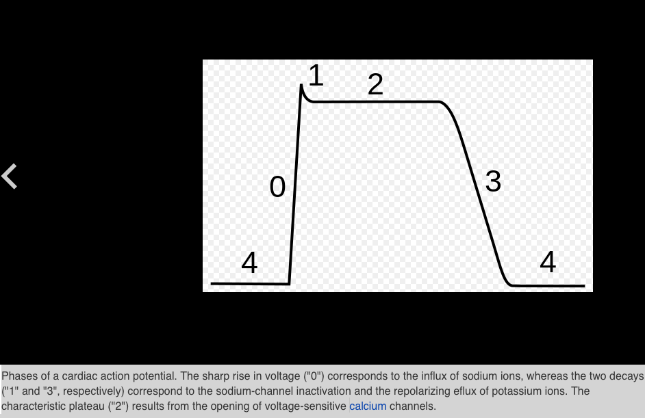
Phases of a cardiac action potential. The sharp rise in voltage ("0") corresponds to the influx of sodium ions, whereas the two decays ("1" and "3", respectively) correspond to the sodium-channel inactivation and the repolarizing eflux of potassium ions. The characteristic plateau ("2") results from the opening of voltage-sensitive calciumchannels.The cardiac action potential differs from the neuronal action potential by having an extended plateau, in which the membrane is held at a high voltage for a few hundred milliseconds prior to being repolarized by the potassium current as usual.[ai] This plateau is due to the action of slower calcium channels opening and holding the membrane voltage near their equilibrium potential even after the sodium channels have inactivated.
The cardiac action potential plays an important role in coordinating the contraction of the heart.[ai] The cardiac cells of the sinoatrial node provide the pacemaker potential that synchronizes the heart. The action potentials of those cells propagate to and through the atrioventricular node (AV node), which is normally the only conduction pathway between the atria and the ventricles. Action potentials from the AV node travel through the bundle of His and thence to the Purkinje fibers.[note 2] Conversely, anomalies in the cardiac action potential—whether due to a congenital mutation or injury—can lead to human pathologies, especially arrhythmias.[ai] Several anti-arrhythmia drugs act on the cardiac action potential, such as quinidine, lidocaine, beta blockers, and verapamil.[aj]
More detailsPhases of a cardiac action potential. The sharp rise in voltage ("0") corresponds to the influx of sodium ions, whereas the two decays ("1" and "3", respectively) correspond to the sodium-channel inactivation and the repolarizing eflux of potassium ions. The characteristic plateau ("2") results from the opening of voltage-sensitive calcium channels.
PNG: Quasar SVG: Mnokel (talk) - This file was derived from: Action potential.png
Basic ventricular myocyte action potential (with labels). 0: depolarization: voltage-gated sodium channels open allowing influx of sodium ions 1: initial repolarization: inactivation of voltage-gated sodium channels; voltage-gated potassium channels begin to open allowing minor efflux of potassium ions 2: plateau: voltage-gated calcium channels open allowing influx of calcium ions triggering calcium release from sarcoplasmic reticulum which results in myocyte contraction 3: rapid repolarization: voltage-gated slow potassium channels open allowing major efflux of potassium ions; voltage-gated calcium channels close 4: resting potential: potassium channels allow for potassium permeability
Permission detailsI, the copyright holder of this work, hereby publish it under the following licenses:
This file is licensed under the Creative Commons Attribution-Share Alike 3.0 Unported license. Subject to disclaimers.
You are free:
Muscular action potentials[edit]Main articles: Neuromuscular junction and Muscle contractionThe action potential in a normal skeletal muscle cell is similar to the action potential in neurons.[46] Action potentials result from the depolarization of the cell membrane (the sarcolemma), which opens voltage-sensitive sodium channels; these become inactivated and the membrane is repolarized through the outward current of potassium ions. The resting potential prior to the action potential is typically −90mV, somewhat more negative than typical neurons. The muscle action potential lasts roughly 2–4 ms, the absolute refractory period is roughly 1–3 ms, and the conduction velocity along the muscle is roughly 5 m/s. The action potential releases calcium ions that free up the tropomyosin and allow the muscle to contract. Muscle action potentials are provoked by the arrival of a pre-synaptic neuronal action potential at the neuromuscular junction, which is a common target for neurotoxins.[ag]
Phases of a cardiac action potential. The sharp rise in voltage ("0") corresponds to the influx of sodium ions, whereas the two decays ("1" and "3", respectively) correspond to the sodium-channel inactivation and the repolarizing eflux of potassium ions. The characteristic plateau ("2") results from the opening of voltage-sensitive calciumchannels.The cardiac action potential differs from the neuronal action potential by having an extended plateau, in which the membrane is held at a high voltage for a few hundred milliseconds prior to being repolarized by the potassium current as usual.[ai] This plateau is due to the action of slower calcium channels opening and holding the membrane voltage near their equilibrium potential even after the sodium channels have inactivated.
The cardiac action potential plays an important role in coordinating the contraction of the heart.[ai] The cardiac cells of the sinoatrial node provide the pacemaker potential that synchronizes the heart. The action potentials of those cells propagate to and through the atrioventricular node (AV node), which is normally the only conduction pathway between the atria and the ventricles. Action potentials from the AV node travel through the bundle of His and thence to the Purkinje fibers.[note 2] Conversely, anomalies in the cardiac action potential—whether due to a congenital mutation or injury—can lead to human pathologies, especially arrhythmias.[ai] Several anti-arrhythmia drugs act on the cardiac action potential, such as quinidine, lidocaine, beta blockers, and verapamil.[aj]
More detailsPhases of a cardiac action potential. The sharp rise in voltage ("0") corresponds to the influx of sodium ions, whereas the two decays ("1" and "3", respectively) correspond to the sodium-channel inactivation and the repolarizing eflux of potassium ions. The characteristic plateau ("2") results from the opening of voltage-sensitive calcium channels.
PNG: Quasar SVG: Mnokel (talk) - This file was derived from: Action potential.png
Basic ventricular myocyte action potential (with labels). 0: depolarization: voltage-gated sodium channels open allowing influx of sodium ions 1: initial repolarization: inactivation of voltage-gated sodium channels; voltage-gated potassium channels begin to open allowing minor efflux of potassium ions 2: plateau: voltage-gated calcium channels open allowing influx of calcium ions triggering calcium release from sarcoplasmic reticulum which results in myocyte contraction 3: rapid repolarization: voltage-gated slow potassium channels open allowing major efflux of potassium ions; voltage-gated calcium channels close 4: resting potential: potassium channels allow for potassium permeability
Permission detailsI, the copyright holder of this work, hereby publish it under the following licenses:
This file is licensed under the Creative Commons Attribution-Share Alike 3.0 Unported license. Subject to disclaimers.
You are free:
- to share – to copy, distribute and transmit the work
- to remix – to adapt the workUnder the following conditions:
- attribution – You must attribute the work in the manner specified by the author or licensor (but not in any way that suggests that they endorse you or your use of the work).
- share alike – If you alter, transform, or build upon this work, you may distribute the resulting work only under the same or similar license to this one.This licensing tag was added to this file as part of the GFDL licensing update.Permission is granted to copy, distribute and/or modify this document under the terms of the GNU Free Documentation License, Version 1.2 or any later version published by the Free Software Foundation; with no Invariant Sections, no Front-Cover Texts, and no Back-Cover Texts. A copy of the license is included in the section entitled GNU Free Documentation License. Subject to disclaimers.
- CC BY-SA 3.0hide terms
- File:Ventricular myocyte action potential.svg
- Uploaded by Hazmat2
- Created: 7 August 2009
Muscular action potentials[edit]Main articles: Neuromuscular junction and Muscle contractionThe action potential in a normal skeletal muscle cell is similar to the action potential in neurons.[46] Action potentials result from the depolarization of the cell membrane (the sarcolemma), which opens voltage-sensitive sodium channels; these become inactivated and the membrane is repolarized through the outward current of potassium ions. The resting potential prior to the action potential is typically −90mV, somewhat more negative than typical neurons. The muscle action potential lasts roughly 2–4 ms, the absolute refractory period is roughly 1–3 ms, and the conduction velocity along the muscle is roughly 5 m/s. The action potential releases calcium ions that free up the tropomyosin and allow the muscle to contract. Muscle action potentials are provoked by the arrival of a pre-synaptic neuronal action potential at the neuromuscular junction, which is a common target for neurotoxins.[ag]
 RSS Feed
